Documentation
Description of Reports, Records, Images, and Analyses
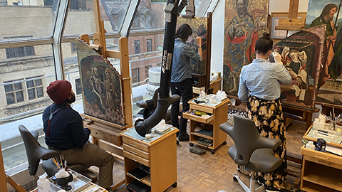
The development of the Kress Program website has been a significant undertaking involving compiling, digitizing, organizing, and editing over 30 years of reports, images, analyses, and research materials. The digitization of these files ensures their preservation and allows for us to share them with wider audiences.
Documentation standards and technologies have evolved over time. Below, documentation categories are outlined, citing the technology and equipment(s) used and the process by which they were acquired.
Catalogue Entries
From 1964-1977, the Kress Foundation sponsored the publication of nine illustrated volumes detailing the entire collection. The Complete Catalogue of the Samuel H. Kress Collection: Italian Paintings XIII-XV Century, Italian Paintings XV-XVI Century, and Italian Paintings XVI-XVIII Century written by Fern Rusk Shapley, include artist information, provenance, art historical scholarship, and related references on every Italian painting in the collection. European Paintings Excluding Italian by Colin Eisler completes the four volumes devoted to Kress paintings. These volumes are available for download and online browsing.
Federico Zeri published many additional opinions in a 1967 Burlington Magazine review that have been incorporated as addenda. While most portions of the catalogues remain authoritative and current, study and treatment of paintings over the years have inevitably led to changes in attribution. Some entries have been updated by Dianne Dwyer Modestini and Shan Kuang to reflect current scholarship and research.
Examination Reports
Examination reports include information about the condition of the painting, including discussions of previous conservation treatments, new technical analyses, and the artist’s materials and techniques. The standard content and format have shifted over the years, resulting in longer examination reports. Some older reports were type-written before the development of word processing and others were derived from hand written notes. As part of this project, all reports were scanned, transcribed, edited, and standardized.
Treatment Reports
Treatment reports describe the methods and materials used during the conservation treatment of the paintings. These reports often include field-specific nomenclature and trade names for materials which may be unfamiliar to a general audience. Please refer to our Glossary for definitions and images.
Photographic Documentation
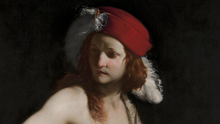
Photographing a painting before treatment, during treatment, and after treatment is an essential aspect of conservation treatment. These photographs document the condition of the painting, what treatment(s) has been carried out, and where the restorations are present to inform conservators and scholars. The following photographs may be present within an object file:
- Before treatment: the appearance before the treatment had taken place. Deteriorated yellowed varnishes and discolored restorations are often notable.
- During treatment: any intermediate appearance of the painting while the treatment is ongoing. For example, they can capture striking varnish removal transformations during treatment or be taken to show the progress of retouching.
- Cleaned state: the appearance of the painting after cleaning, when the varnish and old restorations had been removed. In this state, it is possible to study the condition of the original painting.
- After treatment: the appearance after the completion of retouching and the application of final varnish coatings.
- Ultraviolet (UV): photographs shot in ultraviolet light can provide information about surface coatings, reveal the locations of retouchings, and characterize certain pigments that fluoresce in UV.
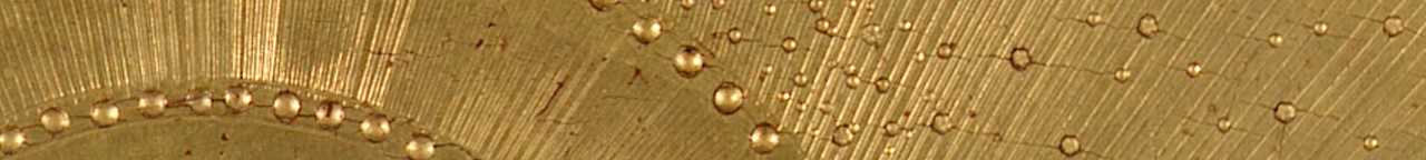
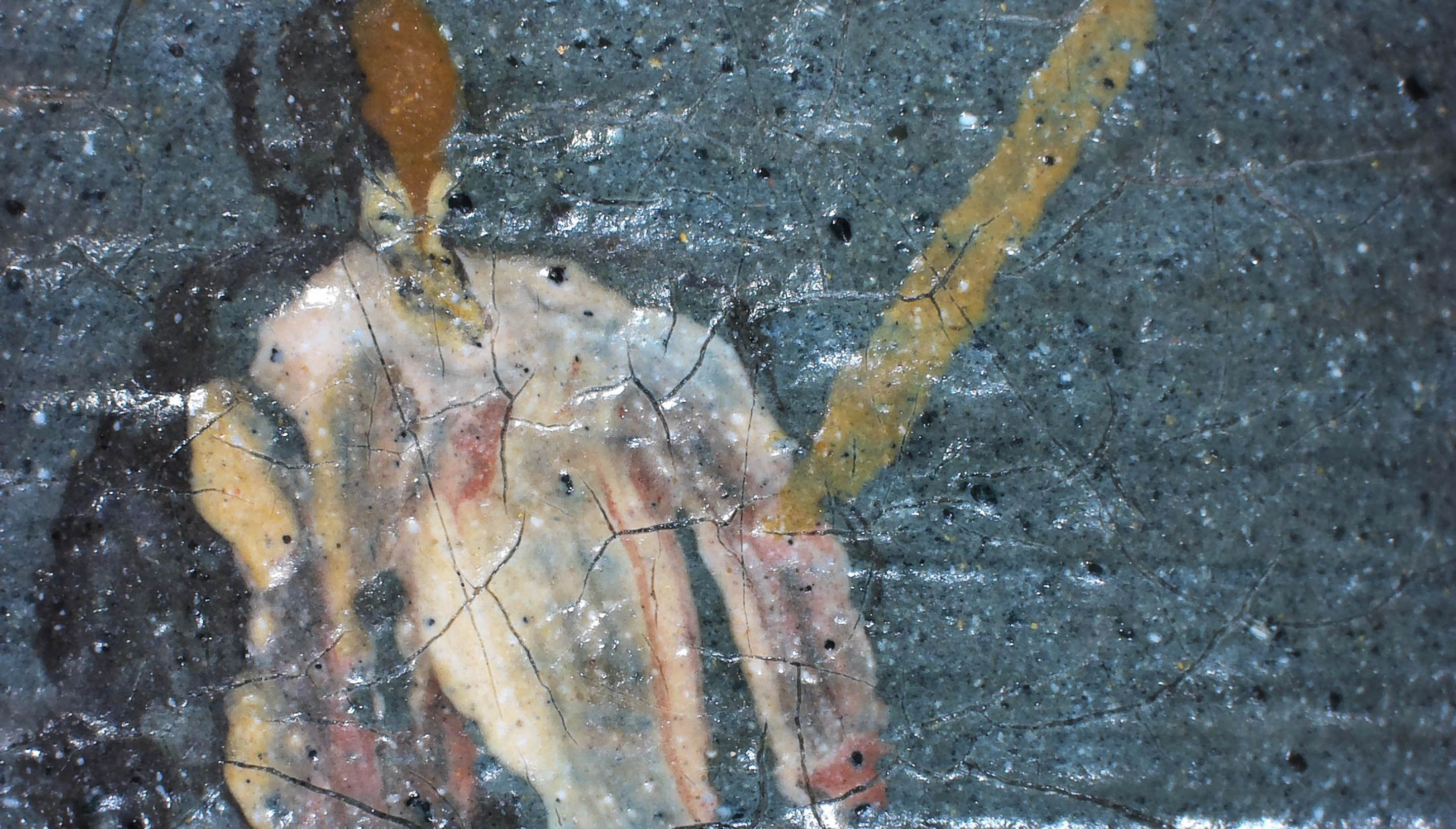
- Photomicrographs: details of the painting photographed with a camera attached to a microscope. Photomicrographs can document in detail surface topographies, pigment mixtures, paint layering techniques, and material condition.
Before 2010, photo documentations were recorded with 8” x 10” color transparencies, 4” x 5” color transparencies, 35mm color slides, and black and white photographs. During this project, these physical records were digitized and edited.
Since the early 2010s, digital photography replaced transparencies and slides. Photographs were taken with either a Nikon D810 camera or a Nikon D700.
Infrared Reflectography
Infrared reflectography is a technique that uses wavelengths in the infrared region of the electromagnetic spectrum to penetrate through opaque paint layers and reveal otherwise invisible elements of the composition. Infrared light is absorbed by carbon-rich materials and reflected back from light-colored elements such as a white preparation. Increasingly sophisticated sensors using the full range of the infrared spectrum with higher sensitivity and better resolution are now able to reveal various features of a painting - such as pentimenti and underpainted sketches - even on paintings with a darker ground.
Infrared captures are acquired in small segments as the camera scans the surface of a painting. Only the earliest reflectograms were collected with a Hamamatsu Vidicon C2400 infrared camera. Most were captured using an InGaAs sensor with 0.9-1.7 µm range (FLIR SC2500-NIR camera and, since 2016, Apollo Camera by Opus Instruments).
X-Radiography
X-rays are short, high energy wavelengths that can penetrate through all the layers of a painting. The image produced, a radiograph, essentially maps the distribution of pigments containing lead, such as lead white, and to a lesser degree other heavy elements such as mercury-based vermilion that x-rays cannot pass through. It can reveal pentimenti or other ways the artist created or altered the composition.
Many of the radiographs on this website are scans of wet process x-rays. The film negatives are stored in an archive at the Conservation Center. During this project, all wet process x-ray plates were scanned and digitized. Since 2016, radiographs are now captured with the Carestream HPX-1 digital system.
X-Ray Fluorescence (XRF)
X-ray fluorescence is a method of noninvasive elemental analysis used to determine the elemental composition of materials. From the data, many pigments can be inferred from the elements present. XRF spectra are occasionally part of Kress object files, and their significance is interpreted in the reports. Performed with Bruker Tracer Handheld XRF.
Cross Sections
Cross sections are tiny samples of the painted layers, embedded in blocks of Bioplast resin, which reveal the layer structure of the painting. Samples are examined with a Leica DM4000B Microscope. Further analysis with Scanning Electron Microscopy with Energy Dispersed Spectroscopy (SEM-EDS) is performed with a Hitachi TM3000 Tabletop Microscope with Bruker Scan Generator. Raman spectroscopy is performed with a Renishaw Raman System 1000, featuring a Leica DM LM microscope and 785nm laser.
Fourier-Transform Infrared Spectroscopy
Fourier-transform infrared spectroscopy is a method of organic analysis employed to analyze the components of a sample. ATR-FTIR is often used to identify surface coatings and binding agents used on a painting. Performed with Bruker Hyperion FTIR microscope interfaced to a Vertex 70 Bruker Optics spectrometer.